We present a mechanical-scan-free method for volumetric imaging of biological tissue. The optical sectioning is provided by structured illumination, and the depth of the imaging plane is varied using an electrically tunable-focus lens. We characterize and evaluate the ability of this axial-scanning mechanism in structured illumination microscopy and demonstrate its ability to perform subcellular resolution imaging in oral mucosa ex vivo. The proposed mechanism can potentially convert any … [Read more...] about Volumetric structured illumination microscopy enabled by a tunable-focus lens
Journal Publications
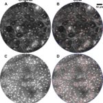
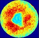
A pulse coupled neural network segmentation algorithm for reflectance confocal images of epithelial tissue
Automatic segmentation of nuclei in reflectance confocal microscopy images is critical for visualization and rapid quantification of nuclear-to-cytoplasmic ratio, a useful indicator of epithelial precancer. Reflectance confocal microscopy can provide three-dimensional imaging of epithelial tissue in vivo with sub-cellular resolution. Changes in nuclear density or nuclear-to-cytoplasmic ratio as a function of depth obtained from confocal images can be used to determine the presence or stage of … [Read more...] about A pulse coupled neural network segmentation algorithm for reflectance confocal images of epithelial tissue
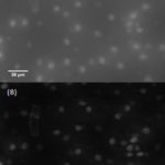
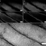
Reflectance confocal endomicroscope with optical axial scanning for in vivo imaging of the oral mucosa
This paper presents the design and evaluation of a reflectance confocal laser endomicroscope using a miniature objective lens within a rigid probe in conjunction with an electrically tunable lens for axial scanning. The miniature lens was characterized alone as well as in the endoscope across a 200 µm axial scan range using the tunable lens. The ability of the confocal endoscope to probe the human oral cavity is demonstrated by imaging of the oral mucosa in vivo. The results indicate that … [Read more...] about Reflectance confocal endomicroscope with optical axial scanning for in vivo imaging of the oral mucosa
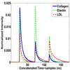
Estimation of the number of fluorescent end-members for quantitative analysis of multispectral FLIM data
Multispectral fluorescence lifetime imaging (m-FLIM) can potentially allow identifying the endogenous fluorophores present in biological tissue. Quantitative description of such data requires estimating the number of components in the sample, their characteristic fluorescent decays, and their relative contributions or abundances. Unfortunately, this inverse problem usually requires prior knowledge about the data, which is seldom available in biomedical applications. This work presents a new … [Read more...] about Estimation of the number of fluorescent end-members for quantitative analysis of multispectral FLIM data
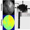
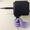
Handheld multispectral fluorescence lifetime imaging system for in vivo applications
There is an increasing interest in the application of fluorescence lifetime imaging (FLIM) for medical diagnosis. Central to the clinical translation of FLIM technology is the development of compact and high-speed clinically compatible systems. We present a handheld probe design consisting of a small maneuverable box fitted with a rigid endoscope, capable of continuous lifetime imaging at multiple emission bands simultaneously. The system was characterized using standard fluorescent dyes. The … [Read more...] about Handheld multispectral fluorescence lifetime imaging system for in vivo applications

Optical axial scanning in confocal microscopy using an electrically tunable lens
This paper presents the use and characterization of an electrically focus tunable lens to perform axial scanning in a confocal microscope. Lateral and axial resolution are characterized over a >250 µm axial scan range. Confocal microscopy using optical axial scanning is demonstrated in epithelial tissue and compared to traditional stage scanning. By enabling rapid axial scanning, minimizing motion artifacts, and reducing mechanical complexity, this technique has potential to enhance in … [Read more...] about Optical axial scanning in confocal microscopy using an electrically tunable lens
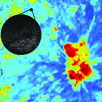
Fluorescence lifetime imaging and reflectance confocal microscopy for multiscale imaging of oral precancer
Optical imaging techniques using a variety of contrast mechanisms are under evaluation for early detection of epithelial precancer; however, tradeoffs in field of view (FOV) and resolution may limit their application. Therefore, we present a multiscale multimodal optical imaging system combining macroscopic biochemical imaging of fluorescence lifetime imaging (FLIM) with subcellular morphologic imaging of reflectance confocal microscopy (RCM). The FLIM module images a 16×16 mm 2 tissue area … [Read more...] about Fluorescence lifetime imaging and reflectance confocal microscopy for multiscale imaging of oral precancer

Chromatic confocal microscopy for multi-depth imaging of epithelial tissue
We present a novel chromatic confocal microscope capable of volumetric reflectance imaging of microstructure in non-transparent tissue. Our design takes advantage of the chromatic aberration of aspheric lenses that are otherwise well corrected. Strong chromatic aberration, generated by multiple aspheres, longitudinally disperses supercontinuum light onto the sample. The backscattered light detected with a spectrometer is therefore wavelength encoded and each spectrum corresponds to a line image. … [Read more...] about Chromatic confocal microscopy for multi-depth imaging of epithelial tissue
Flexible endoscope for continuous in vivo multispectral fluorescence lifetime imaging
Fluorescence lifetime imaging (FLIM) offers a noninvasive approach for characterizing the biochemical composition of biological tissue. There has been an increasing interest in the application of multispectral FLIM for medical diagnosis. Central to the clinical translation of FLIM technology is the development of compact and high-speed endoscopy systems. Unfortunately, the predominant multispectral FLIM approaches suffer from limitations that impede the development of endoscopy systems that are … [Read more...] about Flexible endoscope for continuous in vivo multispectral fluorescence lifetime imaging
Imaging inflammation in mouse colon using a rapid stage-scanning confocal fluorescence microscope
Large area confocal microscopy may provide fast, high-resolution image acquisition for evaluation of tissue in pre-clinical studies with reduced tissue processing in comparison to histology. We present a rapid beam and stage-scanning confocal fluorescence microscope to image cellular and tissue features along the length of the entire excised mouse colon. The beam is scanned at 8,333 lines/sec by a polygon scanning mirror while the specimen is scanned in the orthogonal axis by a motorized … [Read more...] about Imaging inflammation in mouse colon using a rapid stage-scanning confocal fluorescence microscope