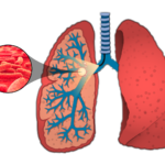
Tuberculosis is one of the deadliest infectious diseases worldwide. New tools to study pathogenesis and monitor subjects in pre-clinical studies to develop treatment regimens are critical for progress. We developed an improved optical system for detecting bacteria in lungs of mice using internal illumination. We present a computational optical model of the full mouse torso to characterize the optical system. Simulated theoretical limits for the lowest detectable bacterial load support the … [Read more...] about Fluorescence modeling of in vivo optical detection of Mycobacterium tuberculosis
Journal Publications

Fabrication and characterization of optical tissue phantoms containing macrostructure
Optical tissue phantoms are essential tools for calibration and characterization of optical imaging systems and validation of theoretical models. This article details a method for phantom fabrication that includes replication of tissue optical properties and three-dimensional tissue structure. … [Read more...] about Fabrication and characterization of optical tissue phantoms containing macrostructure
Optical model of the murine lung to optimize pulmonary illumination
We describe a Monte Carlo model of the mouse torso to optimize illumination of the mouse lung for fluorescence detection of low levels of pulmonary pathogens, specifically Mycobacterium tuberculosis. After validation of the simulation with an internally illuminated optical phantom, the entire mouse torso was simulated to compare external and internal illumination techniques. Measured optical properties of deflated mouse lungs were scaled to mimic the diffusive properties of inflated lungs in … [Read more...] about Optical model of the murine lung to optimize pulmonary illumination

Light scattering by pulmonary alveoli and airway surface liquid using a concentric sphere model
We employ a concentric sphere Mie scattering model to describe light scattering by pulmonary alveoli and airway surface liquid (ASL). Using this layered sphere model, we compare alveolar scattering at different points along the respiratory cycle and observe the effect of ASL thickness on light scattering in the lung. We have also extrapolated the model to investigate alveolar scattering in various animal models of pulmonary disease. This model of pulmonary light scattering can estimate in … [Read more...] about Light scattering by pulmonary alveoli and airway surface liquid using a concentric sphere model
Optically sectioned wide-field fluorescence lifetime imaging microscopy enabled by structured illumination
In this paper, we demonstrate the ability of structured illumination microscopy to enhance the ability of fluorescence lifetime imaging to resolve fluorescence lifetimes in relatively thick samples that possess distinct but spectrally overlapping fluorescent layers. Structured illumination fluorescent lifetime imaging microscopy (SI-FLIM) is shown to be able to accurately reconstruct lifetime values in homogenous fluorophore samples (POPOP, NADH, and FAD) as well as accurately measure … [Read more...] about Optically sectioned wide-field fluorescence lifetime imaging microscopy enabled by structured illumination

Handheld tunable focus confocal microscope utilizing a double-clad fiber coupler for in vivo imaging of oral epithelium
A reflectance confocal endomicroscope with double-clad fiber coupler and electrically tunable focus lens is applied to imaging of the oral mucosa. The instrument is designed to be lightweight and robust for clinical use. The tunable lens allows axial scanning through >250 μm in the epithelium when the probe tip is placed in contact with tissue. Images are acquired at 6.6 frames per second with a field of view diameter up to 850 μm. In vivo imaging of a wide range of normal sites in the oral … [Read more...] about Handheld tunable focus confocal microscope utilizing a double-clad fiber coupler for in vivo imaging of oral epithelium
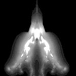
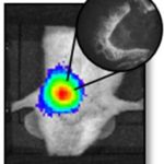
Intravital excitation increases detection sensitivity for pulmonary tuberculosis by whole-body imaging with β-lactamase reporter enzyme fluorescence
Tuberculosis is a pulmonary disease with an especially high mortality rate in immuno‐compromised populations, specifically children and HIV positive individuals. The causative agent, Mycobacterium tuberculosis (Mtb), is a very slow growing and difficult organism to work with, making both diagnosis and development of effective treatments cumbersome. We utilize a fiber‐optic fluorescence microendoscope integrated with a whole‐body imaging system for in vivo Mtb detection. The system exploits an … [Read more...] about Intravital excitation increases detection sensitivity for pulmonary tuberculosis by whole-body imaging with β-lactamase reporter enzyme fluorescence
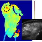
Intravital fluorescence excitation in whole-animal optical imaging
Whole-animal fluorescence imaging with recombinant or fluorescently-tagged pathogens or cells enables real-time analysis of disease progression and treatment response in live animals. Tissue absorption limits penetration of fluorescence excitation light, particularly in the visible wavelength range, resulting in reduced sensitivity to deep targets. Here, we demonstrate the use of an optical fiber bundle to deliver light into the mouse lung to excite fluorescent bacteria, circumventing tissue … [Read more...] about Intravital fluorescence excitation in whole-animal optical imaging
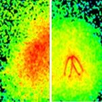
Objective detection of oral carcinoma with multispectral fluorescence lifetime imaging in vivo
Successful early detection and demarcation of oral carcinoma can greatly impact the associated morbidity and mortality rates. Current methods for detection of oral cancer include comprehensive visual examination of the oral cavity, typically followed by tissue biopsy. A noninvasive means to guide the clinician in making a more objective and informed decision toward tissue biopsy can potentially improve the diagnostic yield of this process. To this end, we investigate the potential of … [Read more...] about Objective detection of oral carcinoma with multispectral fluorescence lifetime imaging in vivo
A novel multimodal optical imaging system for early detection of oral cancer
Objectives. Several imaging techniques have been advocated as clinical adjuncts to improve identification of suspicious oral lesions. However, these have not yet shown superior sensitivity or specificity over conventional oral examination techniques. We developed a multimodal, multi-scale optical imaging system that combines macroscopic biochemical imaging of fluorescence lifetime imaging with subcellular morphologic imaging of reflectance confocal microscopy for early detection of oral cancer. … [Read more...] about A novel multimodal optical imaging system for early detection of oral cancer